


Less Segmentation… More Innovation – New Anatomies Added to AI-Powered Simpleware AS Ortho Module
Posted on 8 March 2021 by Celia Butler

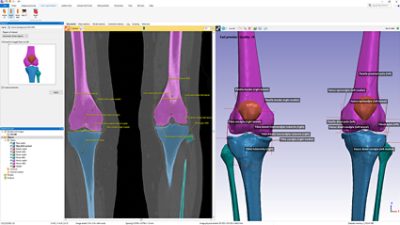
The bones segmented from a lower body CT scan using the Hip CT, Knee CT and Ankle CT tools in Simpleware AS Ortho.
We are delighted to announce the addition of two new anatomy-specific segmentation tools to the Simpleware AS Ortho module: Ankle CT and Knee CT, as well as key updates to the existing Knee MRI tool. This release offers a complete solution for full joint replacement planning by automatically segmenting and landmarking the Hip, Knee and Ankle in just a few clicks.
These new and improved tools join our established Auto Segmenter for Orthopedics which is powered by AI technology using state-of-the-art Machine Learning (ML) algorithms to perform complex and usually time-consuming segmentation and landmarking tasks automatically, and in minutes!
The Simpleware ML algorithms are quick and produce accurate results in a few minutes using standard engineering hardware (including laptops). Running locally, there is no need to upload your confidential patient data to offsite servers for processing – ensuring all your data stays secure within your network.
Our tried and tested algorithms are trained by experts and results are verified by clinical professionals, offering consistency between users that can be scaled up to process large batches of data up to 50 times faster than traditional methods.
Benefits of Using Simpleware AS Ortho
- Fully automated: One-click solution to eliminate hours spent on tedious manual processes.
- Fast and effective: Get results in 1 – 3 minutes on a standard engineering specification laptop.
- Accurate and reliable: Simpleware ML algorithms are trained by experts and verified by clinical professionals.
- Secure: Protect your patient data on your local hardware, avoiding the need to transfer confidential data onto servers outside of your control.
- Consistent and repeatable: Eliminate inconsistencies between users and need for multiple reviews.
- Scalable: Boost your throughput, and efficiently process large numbers of datasets 20 – 50 times faster.
- Less Segmentation - More Innovation: Free up engineering time for more complex and high-value tasks.
New Segmentation and Landmarking Tools
The Ankle CT tool explores a new anatomy as part of Synopsys’ offering to the Orthopedic community. This tool automatically segments the bones in the ankle joint, including the Talus, Calcaneus, Tibia, and Fibula from CT image data. It also automatically places anatomic landmarks on the ankle center, fibular notch as well as the lateral and medial malleolus.

The ankle bones segmented and landmarked from a CT using the new AI-powered Ankle CT tool, part of Simpleware AS Ortho.
The Knee CT tool compliments the existing Knee MRI tool for those working to create knee models from image data. This tool automatically segments the bones in the knee joint including the Femur, Tibia, Fibula, Patella and Fabella if present from CT image data. In addition to the segmentation the tools, this approach also automatically places an extensive range of anatomic landmarks on the bones segmented.
The left and right knees segmented and landmarked from a CT scan using the new AI-powered Knee CT tool, part of Simpleware AS Ortho.
Like all Simpleware AS Ortho tools, Ankle CT and Knee CT include the ability to define and automatically detect specific regions of interest. The tools can also be fully scripted (along with features from Simpleware ScanIP) using Python or C# to integrate into your pipeline, including batch import and processing, with subsequent model export or result sharing – Simpleware AI solutions fit seamlessly within your wider workflows.
Improvements to Existing Tools
We have also been working closely with our customers to improve the existing tools and added a new image weight to the Knee MRI tool. Knee MRI now includes automatic segmentation and landmarking from T2 sagittal MRI data.
As with the existing PD weighted (Cor/Sag) and T1 (Cor), the Knee MRI tool segments the Femur, Tibia, and associated cartilage, Patella and Fibula, as well as automatically placing a range of anatomic landmarks on the bones segmented. This new offering will allow the Knee MRI tool to be used in a wider range of applications by more institutions.
Learn More
Attend our Live Presentation and Demo on March 17, 2021, or watch on-demand after the live session.
Any Questions?
Talk to us about your Orthopedic data and learn about Simpleware AS Ortho and our full range of automated anatomical segmentation and landmarking solutions to find the perfect fit for your application.